46
46
Clinical Insight
P R O B E
• V o l . L I I I • N o . 3 • A p r – J u n 2 0 1 4
ase Discus ion
Geophagia Masquerading as Urolithiasis
Abeygunasekera AM, et al
Saudi J Kidney Dis Transpl
. 2013;24(4):798–799.
Case Presentation
A 17-year-old school girl was admitted to the hospital with
abdominal pain. She was an average student in the class,
with no learning disability. The abdominal examination did
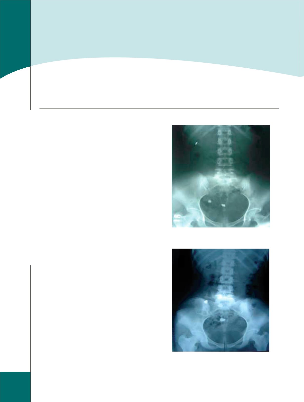
not reveal any signs and the X-ray (kidney, ureter, bladder)
showed 3 opacities in the abdomen, suggestive of a stone in
the right kidney and 2 stones in the lower part of the right
ureter (Figure 1). A diagnosis of renal and ureteric stones
was made and urological opinion was sought. The opacities
had an atypical distribution as the one intended to be in the
lower end of the right ureter was too high up in the X-ray.
The outline of the opacities was too irregular and deformed
to be urinary stones. Therefore, the possibility of stones
in the small intestine was thought of and an abdominal
ultrasonography revealed a normal urinary tract. Another
X-ray obtained 24 h later showed rapid change in the
position of the stones, confirming the presence of stones in
the intestines (Figure 2).
Three days later, the X-ray was normal. When reviewed 6
months later, she was free of abdominal pain or repetition
of geophagia. In this patient, geophagia was not associated
with features of a major psychiatric disorder. By swallowing
stones, the patient has shown a bizarre behavior probably
in a stressful situation, suggesting the possibility of a
fictitious disorder. She did not develop clinical features of
intestinal obstruction or perforation. Medical personnel
should be aware of the possibility of geophagia in patients
with opacities with an atypical distribution in their
abdominal X-rays. Otherwise, patients may be subjected to
unnecessary investigations and delay. The opacities due to
swallowed stones were of varying sizes and densities. Those
with a fictitious disorder do not display major psychiatric or
learning disability, and the diagnosis may be missed if not
associated with a high index of suspicion. Serial radiography
will clinch the diagnosis easily as stones in the intestines
change in position rapidly with time.
Figure 1.
X-ray (kidney, ureter, bladder) Showing 3
Opacities Suggestive of Calculi in the Right Kidney and
Lower Ureter
Figure 2.
X-ray (kidney, ureter, bladder) Taken 24 h Later
Showing Dramatic Change in the Position of Opacities


