28
Clinical insight
P R O B E
• V o l . L I I I • N o . 3 • A p r – J u n 2 0 1 4
colic and of concrements elimination,
real urolithiasis in their families, the
presence of metabolic disorders, and
so forth. During the conduction of the
study, the patients were on free mode
diet and with a fully compensated
renal function. They did not report
any liver disease.
All investigated patients were
hospitalized in the Clinic of the
Department of Urology at the Medical
University, Sofia.
Controls
Fifteen healthy patients who had never
had any urological and hepatic trouble
constituted the control group.
Methods
A 24-h urine collection was obtained
from each patient. During the period
of urine collection, specimens were
refrigerated and aliquots of the 24-h
volume sample were immediately
frozen until analysis. The volume of
urine in every sample was recorded
on completion of the collection and
pH was measured by using a glass
electrode pH meter.
The calcium, oxalate, and other
substances, such as creatinine,
magnesium, phosphorous, uric acid,
were also determined. All substances
were measured by automated known
spectrophotometry and colorimetric
analysis. The urine levels of oxalates
were evaluated by enzymatic method.
Amino acid contents (HA, glycine,
serine, and so forth) of the collected
samples were determined using a high-
performance liquid chromatography
(HPLC) 1050 instrument
(Hewlett Packard), coupled to a
fluorescence detector. Ethyl alcohol
was added to urine specimens to
allow the precipitation of proteins
and the extraction of free amino
acids. An automated precolumn
orthophtaldehyde derivation
procedure was employed. Separations
were done using a reversed-phase
column (Waters Corp). Amino acid
concentration of the samples was
determined in comparison to the
values obtained from a standard curve
prepared for each amino acid.
Statistical analysis
Statistical analysis of the data obtained
from both the stone formers (SF) and
from the control group was performed
using Student
t
test, to establish the
significance of the difference between
mean values. All results were expressed
as a mean ± SEM and differences were
considered significant if
P
< .05.
Dissolution of CaOX
concrements with hippuric
acid: in vitro experiments
Instrumental techniques
The experiments on the kinetics of
the dissolution of CaOX renal calculi
were performed in Jena glass round
bottom flasks thermostated at 25°C.
The volume of the studied solution
was 1000 mL and it was stirred (~ 200
rpm) by an electromagnetic stirrer.
The Archimedean weight G(t) of the
samples of CaOX calculi, put in a
platinum net basket and suspended to
a torsion balance, was continuously
measured with a sensitivity of ± 0.5
mg. The CaOX calculi used had
been formed in the urinary tract
and eliminated spontaneously by the
patients. The calculi were selected to
have a weight of 100 to 200 mg and to
be of identical mineral composition,
mainly CaC
2
O
4
• 2H
2
O (weddellite).
The composition of the calculi was
checked by polarized light microscopy
and thermogravimetry . We employed
2 different types of aqueous solutions,
mimicking urine, with our solvent
(HA) introduced in several different
concentrations.
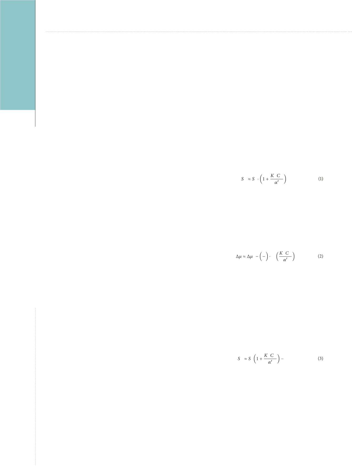
The physico-chemical formalism of
the kinetics of dissolution of kidney
stones has been developed, in details,
in our paper. One can see that simple
formulae can be obtained, describing
the effect of complex forming agents
(present in the solution at various
concentrations) on supersaturation,
solubility, and growth velocity of
CaOX crystals growing or dissolving
in a solution, resembling human urine.
It can be shown that if we introduce
an increasing concentration
C
H
(eg,
HA) of a Ca
2+
-binding complex
forming agent, having a solubility
constant
K
H
into the solution, a linear
dependence between the solubility
S
H
and
C
H
for Ca
2+
>> C
2
O
4
2−
can be
predicted by
where
a
´ is the
a
-factor in the absence
of the complex forming agent H (here
indicating hippuric acid).
Thus, the dependence of the
supersaturation Δ
m
on
C
H
for the
physiologically significant case
Ca
2+
>> C
2
O
4
2−
, determining the
supersaturation in urine is
where Δ
m
0
is the supersaturation in
respect to the CaOX precipitation
without HA added.
It is also of significant interest that in
the case of the dissolution of CaOX
concrements in the presence of a fixed
initial concentration of CaOX (or
which in the case of Ca
2+
>> C
2
O
4
2−
is
the same in the presence of constant
concentration C
0
*
of oxalic anions
Ca
2+
we have to rewrite (1) as follows:
Thus, a plot of
S
H
versus
C
H
should
result in a straight line with a slope of
−
S
•
K
H
/
a
´ cutting from the ordinate
axis a segment
S
H
(0) =
S
−
C
0
*
. In
this way, both
S
H
and
K
H
can be
determined at a known value of
a
,
a
´
in human urine is approximately 2).
Atanassova SS, et al.
Regulation of Supersaturation in Calcium Oxalate Lithiasis
H o
H H
o
1
2
ln
H H
H o
H H
C
o
*


